Home » Without Label » Anatomy Of Musckes Sndctendons : 1 - *the origin, insertion, and belly.* a muscle's origin is where a tendon attaches it to the *less* movable bone.
Anatomy Of Musckes Sndctendons : 1 - *the origin, insertion, and belly.* a muscle's origin is where a tendon attaches it to the *less* movable bone.
Anatomy Of Musckes Sndctendons : 1 - *the origin, insertion, and belly.* a muscle's origin is where a tendon attaches it to the *less* movable bone.. The tendon is firmly connected to muscle fibres at one end and to components of the bone at its other end. Tendons are thick bands of tissue that connect muscles to bones. Originates from the medial and lateral surfaces of the humeral shaft and inserts into the ulnar tuberosity, just distal to the elbow joint. Lying exposed between the protective bones of the superiorly located ribs and the inferiorly located pelvic girdle, the muscles of this region play a critical role in protecting the. The muscles of the abdomen, lower back, and pelvis are separated from those of the chest by the muscular wall of the diaphragm, the critical breathing muscle.
The image below shows the bones of the hand from the back side. Muscles of the hips and thighs. Find the best weight lifting exercises that target each muscle or groups of muscles. Lesson on the anatomy of the forearm: The tendon is firmly connected to muscle fibres at one end and to components of the bone at its other end.
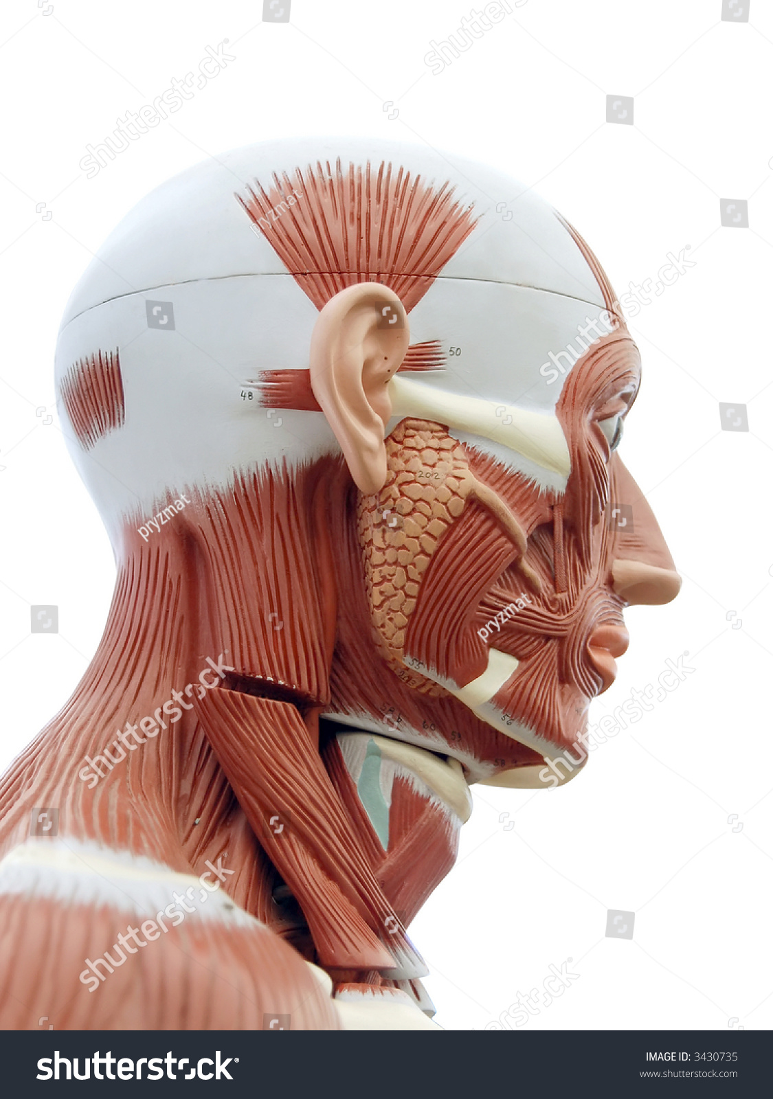
Human Anatomy Structure Head Muscles Tendons Stock Photo Edit Now 3430735 from image.shutterstock.com The calf muscles (gastrocnemius and soleus), which are connected to the calcaneus via the achilles tendon. When the muscle contracts, the tendons are pulled, and the bone is moved. There are three layers of gluteal muscles on the posterior hips, just like there are three layers of muscles in the abdominal trunk. Skeletal muscles are attached to bones by tendons and can be as long as 30 cm, although they are usually 2 to 3 cm in length. The tightening and relaxing of the calf muscles enables the ankle to bend downward and upward. The fleshy, thick part of the muscle is called its belly. It forms the floor of the cubital fossa. Ligaments connect two or more bones together and help stabilize joints.
This important tendon in the back of the calf and ankle stores the elastic.
By connecting our rigid bones to our powerful muscles, tendons allow us to move. Webmd's knee anatomy page provides a detailed image and definition of the knee and its parts including ligaments, bones, and muscles. Human anatomy and physiology lab (bsb 141) module 9: The shoulder girdle includes three bones—the scapula, clavicle and humerus. Ligaments and tendons are fibrous bands of connective tissue that attach to bone. This important tendon in the back of the calf and ankle stores the elastic. Movement occurs when our muscles pull on our bones, relocating them. Lesson on the anatomy of the forearm: A tendon connects the muscle to the bone. The right scapula from the front and back side. Four muscles and their attached tendons make up the rotator cuff. The human hand is made up of the wrist, palm, and fingers and consists of 27 bones, 27 joints, 34 muscles, over 100 ligaments and tendons, and many blood vessels and nerves. There are three layers of gluteal muscles on the posterior hips, just like there are three layers of muscles in the abdominal trunk.
*the origin, insertion, and belly.* a muscle's origin is where a tendon attaches it to the *less* movable bone. Each of them aids in a specific motion of your shoulder. Tendons vary in size and are somewhat elastic and attach bones to muscles. Extensor carpi radialis brevis extensor carpi radialis longus Muscle anatomy dictionary 12 photos of the muscle anatomy dictionary muscle anatomy dictionary, human muscles, muscle anatomy dictionary.
Anatomy Of Knee from ix-cdn.b2e5.com The majority of muscles in the leg are considered long muscles, in that they stretch great distances. The muscles you probably know the best are your glutes. There are three layers of gluteal muscles on the posterior hips, just like there are three layers of muscles in the abdominal trunk. The calf muscles (gastrocnemius and soleus), which are connected to the calcaneus via the achilles tendon. Lying exposed between the protective bones of the superiorly located ribs and the inferiorly located pelvic girdle, the muscles of this region play a critical role in protecting the. The hands enable us to perform many of our daily activities such as driving, writing and cooking. It forms the floor of the cubital fossa. The primary function of the shoulder girdle is to give strength and range of motion to the arm.
The brachialis muscle lies deep to the biceps brachii, and is found more distally than the other muscles of the arm.
Originates from the medial and lateral surfaces of the humeral shaft and inserts into the ulnar tuberosity, just distal to the elbow joint. The knee joint is most significantly affected by two major muscle groups: Anatomy muscles exam questions 12 photos of the anatomy muscles exam questions anatomy muscle test questions, anatomy muscles exam questions, anatomy muscular system test questions, human muscles, anatomy muscle test questions, anatomy muscles exam questions, anatomy muscular system test questions Tendons are the connective tissues that transmit the mechanical force of muscle contraction to the bones; Tendons attach muscle to bone. The posterior tibialis muscle, which supports the arch of the foot and enables the foot to turn. Ligaments and tendons are fibrous bands of connective tissue that attach to bone. The shoulder is not a single joint, but a complex arrangement of bones, ligaments, muscles, and tendons that is better called the shoulder girdle. Human anatomy and physiology lab (bsb 141) module 9: Tendon, tissue that attaches a muscle to other body parts, usually bones. This is lesson 1 on the anatomy of the forearm. This video also provides you with a. In this lesson, we look at the muscle.
Movement occurs when our muscles pull on our bones, relocating them. It forms the floor of the cubital fossa. The quad muscles— which form the meaty mass on the front of your thighs — are among your strongest muscle groups, and play a critical role in athletic activities. This video also provides you with a. Related posts of diagram of shoulder muscles and tendons anatomy muscles exam questions.
Human Male Body Anatomy Illustration Of A Human Back With Visible Muscles Stock Illustration Illustration Of Black Body 111007639 from thumbs.dreamstime.com The quad muscles— which form the meaty mass on the front of your thighs — are among your strongest muscle groups, and play a critical role in athletic activities. As these muscles contract and relax, they move skeletal bones to create movement of the body. Together, these muscles straighten your knee, stabilize your knee joint, assist in flexing your hip (drawing your knee towards your chest), and help absorb force when you land after jumping or leaping. The calf muscles (gastrocnemius and soleus), which are connected to the calcaneus via the achilles tendon. The image below shows the bones of the hand from the back side. Tendons attach muscle to bone. Originates from the medial and lateral surfaces of the humeral shaft and inserts into the ulnar tuberosity, just distal to the elbow joint. The shoulder is not a single joint, but a complex arrangement of bones, ligaments, muscles, and tendons that is better called the shoulder girdle.
The knee joint is most significantly affected by two major muscle groups:
The human hand is made up of the wrist, palm, and fingers and consists of 27 bones, 27 joints, 34 muscles, over 100 ligaments and tendons, and many blood vessels and nerves. Muscles of the hips and thighs. The image below shows the bones of the hand from the back side. The primary function of the shoulder girdle is to give strength and range of motion to the arm. Tendons vary in size and are somewhat elastic and attach bones to muscles. When the muscle contracts, the tendons are pulled, and the bone is moved. On the other hand, the insertion is where a tendon attaches that muscle to the *more* movable bone. The shoulder is not a single joint, but a complex arrangement of bones, ligaments, muscles, and tendons that is better called the shoulder girdle. The tendon is firmly connected to muscle fibres at one end and to components of the bone at its other end. This is lesson 1 on the anatomy of the forearm. Related posts of diagram of shoulder muscles and tendons anatomy muscles exam questions. Related posts of muscles and tendons of the leg muscle anatomy dictionary. Webmd's knee anatomy page provides a detailed image and definition of the knee and its parts including ligaments, bones, and muscles.